| From Embryo to Tumor: Trace Claudin-6 with MS Validated Antibodies |
|
Claudin-6 (MSVA-916R) Therapeutic target protein |
| Datasheet |
|
Biology behind
|
| Claudin-6 (CLDN6) is one of 26 members of the claudin family of membrane proteins that are crucial components of tight junctions, regulating paracellular permeability and maintaining cell polarity. Claudin-6 is coded by the CLDN6 gene on chromosome 16p13.3. Its molecular weight is approximately 23 kDa. CLDN6 is one of the earliest proteins expressed in embryonic stem cells. It is expressed in specific fetal tissues such as the kidney, lung, pancreas, and the stomach, but it is not expressed in the corresponding adult tissues. That knockout mice lacking mouse homolog CLDN6 do not exhibit an aberrant phenotype indicates that CLDN6 is dispensable for normal development and tissue homeostasis. While CLDN6 expression is largely lacking on normal tissues, it is highly expressed in several cancers. Therefore, CLDN6 is considered a potential therapeutic target. Studies evaluating CLDN6 as a target for monoclonal antibodies, antibody-drug conjugates, bispecific antibodies, and CAR-T cells are underway. |
|
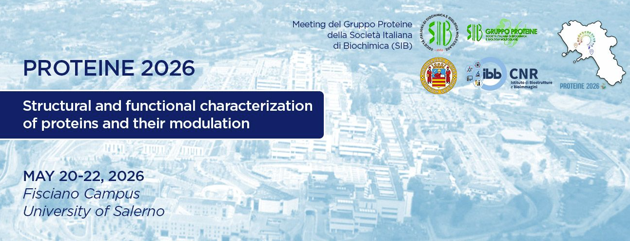
CLDN6 staining in normal tissues is rare. It is strongest in cytotrophoblast cells of the first trimester placenta. A weak to moderate membranous CLDN6 can also occur in few chorion cells, few intercalated ducts and/or acinar cells of the pancreas, few epithelial cells of the adenohypophysis, and in few tubular or collecting duct cells of the kidney.
|
 |
Breast - Absence of CLDN6 staining of epithelial cells in this sample |
| Normal Tissue Gallery |
|
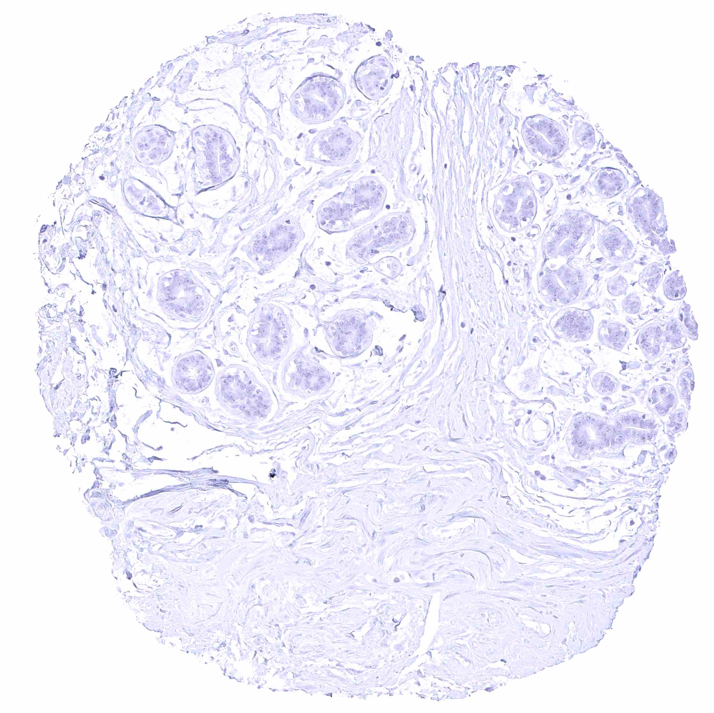
CLDN6 expression is particularly frequent in testicular germ cell tumors as well as in ovarian and endometrial cancer but it also occurs in various other tumor entities.
|
| Testis - seminoma with strong CLDN6 immunostaining of all tumor cells |  |
| Cancer Tissue Gallery |
| Protocol Recommendations |
|
IHC users have different preferences on how the stains should look like. Some prefer high staining intensity of the target stain and even accept some background. Others favor absolute specificity and lighter target stains. Factors that invariably lead to more intense staining include higher concentration of the antibody and visualization tools, longer incubation time, higher temperature during incubation, higher temperature and longer duration of the heat induced epitope retrieval (slide pretreatment). The impact of the pH during slide pretreatment has variable effects and depends on the antibody and the target protein. Manual protocol: Freshly cut sections should be used (less than 10 days between cutting and staining). Heat-induced antigen retrieval for 5 minutes in an autoclave at 121°C in pH 7,8 Target Retrieval Solution buffer. Apply MSVA-916R at a dilution of 1:150 at 37°C for 60 minutes. Visualization of bound antibody by the EnVision Kit (Dako, Agilent) according to the manufacturer’s directions. |
Come and meet us in Fisciano!