

FLuoEVs as a Robust Positive Control for EV Uptake Assays
| Introduction |
|
The study of EV internalization is essential for understanding EV-mediated intercellular communication and therapeutic cargo delivery. Reliable interpretation of uptake experiments requires appropriate controls to distinguish true intracellular EV signal from fluorescence artefacts arising from free dye, nonspecific membrane staining, or imaging misconfiguration. The inclusion of a standardized positive control is therefore critical for reliable assay validation.
|
| Material and Methods |
|
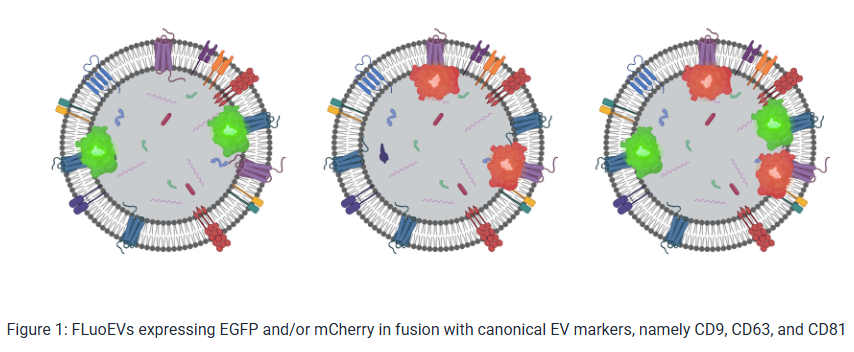
FF-derived EVs were fluorescently labeled using a commercial EV Labeling kit, according to the manufacturer’s instructions. To ensure efficient removal of unbound dye, labelled samples were purified using miniPURE-EVs spin columns (HansaBioMed Life Sciences). To evaluate possible dye-derived artefacts, DPBS was used as dye-only negative control. FluoEVs CD63-EGFP EVs were used directly as a positive control without additional labelling or purification steps. The granulosa-like tumor cell line KGN was used as a cellular model for uptake experiments. A total of 70,000 cells were seeded onto 13 mm glass coverslips and cultured for 24 h prior to treatment. Cells were then incubated for 6 h with 108 FF EVs, 108 FluoEVs, or dye-treated and column-purified DPBS (negative control). Following incubation, cells were washed 3× with DPBS and fixed with 4% paraformaldehyde for 10 min. Cells were then permeabilized using 0.5% Triton X-100 for 5 min and blocked with 5% BSA for 1 h. To visualize cellular architecture, samples were incubated overnight at 4°C with mouse anti-tubulin antibody, followed by a 1 h incubation with DyLight 405 secondary antibody. Coverslips were mounted using ProLong Gold Antifade Mountant. Imaging was performed using a Zeiss LSM 900 microscope equipped with a Plan-Apochromat 63×/1.4 oil immersion objective. Serial z-stacks were acquired using bidirectional scanning with line averaging to ensure high signal-to-noise ratio. All images were captured under identical laser intensity and detector gain settings across all conditions to ensure direct comparability. |
| Results |
|
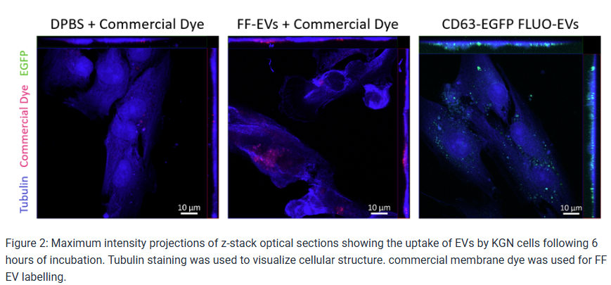
KGN cells treated with CD63-EGFP FluoEVs exhibited bright and well-defined intracellular fluorescent puncta distributed throughout the cytoplasmic compartment. Fluorescence was consistently detectable In contrast, cells treated with membrane dye-labelled FF EVs displayed weaker and more diffuse fluorescence compared to FluoEV-treated cells. Cells exposed to dye-treated and column-purified DPBS showed no detectable intracellular fluorescence under identical imaging settings, confirming effective removal of unbound dye and absence of dye-derived artefacts. The strong and reproducible EGFP signal provided by FluoEVs facilitated reliable calibration of confocal imaging parameters and validation of uptake assay conditions prior to analysis of experimental EV samples. |
| Conclusion |
|
